They have a complex, adaptive network of protective blood vessels that make the structures in our necks look puny–a network that researchers have now dissected, mapped and illustrated for the first time.
“Until now, brain imaging specialists like me who deal with human injuries caused by trauma to arteries in the head and neck have always been puzzled as to why rapid, twisting head movements did not leave thousands of owls lying dead on the forest floor from stroke,” said Dr. Philippe Gailloud, an interventional neuroradiologist at Johns Hopkins and a senior researcher on the study, in a statement. A poster depicting these findings won first place in the 2012 International Science and Engineering Visualization Challenge, the journal Science announced yesterday.
The carotid and vertebral arteries in the neck of most animals, including owls and humans, are delicate and fragile structures. They’re highly susceptible to minor tears and stretches of vessel linings. In humans, such injuries can be common: whiplash sustained in a car accident, a back-and-forth jarring roller coaster ride or even a chiropractic maneuver gone wrong. But they’re also dangerous. Blood vessel tears caused by sudden twisting motions produce clots that can break off, sometimes causing an embolism or stroke that could prove fatal.
Owls, on the other hand, can rotate their necks up to 270 degrees in either direction without damaging the vessels running below their heads, and they can do it without cutting off blood supply to their brains.
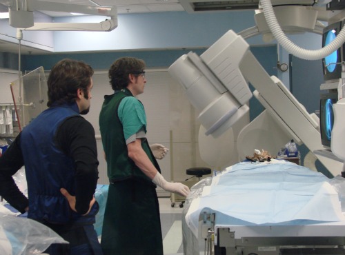
Researchers
Philippe Gailloud (right) and Fabian de Kok-Mercado (left) examine the
bone and vascular structure of an owl that died of natural causes. Photo
courtesy of Johns Hopkins
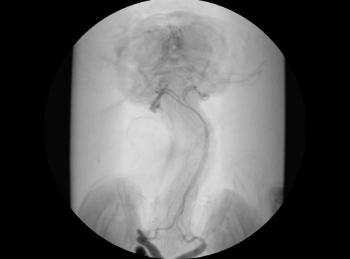
When researchers injected dye into the owls’ arteries to mimic blood flow and then manually turned the birds’ heads, they saw mechanisms at play that contrasted greatly with humans’ head-turning ability. Blood vessels at the base of the owls’ heads, just below the jawbone, kept expanding as more of the dye flowed in. Eventually, the fluid pooled into tiny reservoirs. Our arteries tend to get smaller during head rotations and don’t balloon in the same way.
Dye
injected into deceased owls’ blood vessels pool in tiny reservoirs as
their heads are rotated manually, a feature that allows for
uninterrupted blood flow to the brain. Image courtesy of Johns Hopkins
But these silent hunters’ head-on-a-swivel ability continued to be more complex, researchers found. In owls’ necks, one of the major arteries feeding the brain passes through bony holes in the birds’ vertebrae. These hollow cavities, known as the transverse foraminae, were ten times bigger in diameter than the artery passing through it. The researchers say the roomy extra space creates multiple air pockets that cushion the artery and allow it to travel safely during twisting motions.
“In humans, the vertebral artery really hugs the hollow cavities in the neck. But this is not the case in owls, whose structures are specially adapted to allow for greater arterial flexibility and movement,” said lead researcher Fabian de Kok-Mercado in the statement. De Kok-Mercado is a medical illustrator at Howard Hughes Medical Institute in Maryland.
This adaptation appeared in 12 of the 14 vertebrae in the owls’ necks. The vertebral arteries entered their necks higher up than in other birds, introduced at the 12th vertebrae (when counted from the top) instead of the 14th, which gives the vessels more slack and room to breathe. Small vessel connections between the carotid and vertebral arteries, called anastomoses, let blood flow uninterrupted to the brain, even when owls’ necks were contorted into the most extreme twists and turns.
“Our in-depth study of owl anatomy resolves one of the many interesting neurovascular medical mysteries of how owls have adapted to handle extreme head rotations,” de Kok-Mercado said.
Up next for the team is studying hawk anatomy to find out if other bird species possess owls’ adaptive features for looking far left and right.


No comments:
Post a Comment